Differentiable Electron Microscopy Simulation: Methods and Applications for Visualization
At a glance
- Introduces DiffTEM, a scalable differentiable simulator for electron-microscopy micrographs.
- Models deterministic image formation and stochastic noise in one differentiable pipeline.
- Demonstrates automatic detector-parameter estimation and real-microscopy denoising applications.
Motivation
Electron-microscopy data are expensive to acquire, noisy, and difficult to annotate. Faithful simulation can supply controlled ground truth, but conventional simulators are often too slow or too rigid to support optimization and learning.
Method
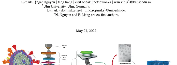
DiffTEM simulates micrographs from atomistic models at a scale suitable for many viral particles. Both the deterministic image-formation stages and the stochastic signal-and-noise stages are differentiable, allowing gradients to flow from an image-space objective back to simulation and detector parameters.
Evaluation
The paper demonstrates two uses of differentiability: estimating detector parameters automatically and using simulated data to support denoising of real microscopy observations. It also evaluates the simulator’s visual plausibility and scalability relative to previous simulation approaches.
Limitations
A simulator remains an approximation of a complex physical acquisition process. Its usefulness depends on how well the modeled specimen, microscope, and noise assumptions match the data to which it is applied.